Foot Muscles Mri - Mri Of The Left Foot In A Normal Patient For Comparison Coronal Download Scientific Diagram / Magnetic resonance imaging, otherwise known as mri, uses a combination of magnetic fields and radio waves to take images of the internal structures of your body.
Foot Muscles Mri - Mri Of The Left Foot In A Normal Patient For Comparison Coronal Download Scientific Diagram / Magnetic resonance imaging, otherwise known as mri, uses a combination of magnetic fields and radio waves to take images of the internal structures of your body.. The traditional full body mri can cost up to $3,500 limiting patients who need the imaging to get a full and proper diagnosis. This imaging technique assesses the ligaments and tendons, neurovascular structures (tarsal tunnel and plantar fascia), and the osseous structures(19). Denervation changes in muscles early. The aim of this review is to provide the reader with a comprehensive overview of the magnetic resonance imaging (mri) characteristics of the most common benign and malignant soft tissue neoplasms which occur around the foot and ankle. It flexes and extends the foot, ankle, and knee.
23 it can originate as a separate muscle from the fibula or from the peroneus brevis or longus muscles and inserts onto the peroneal tubercle or retrotrochlear eminence of the calcaneus. In addition, an image of all the muscles of the back and plantar part of the foot, all tendons and tendon ligaments, blood vessels and nerves are obtained. Those fibers of the most medial and largest belly are… The majority of soft tissue lesions in the foot and ankle are benign. Muscle anatomy trivia 12 photos of the muscle anatomy trivia muscle anatomy trivia, human muscles, muscle anatomy trivia
The most common ossicle is the os trigonum, which is a prominent unfused apophysis of the lateral tubercle of the talus.
Magnetic resonance imaging, otherwise known as mri, uses a combination of magnetic fields and radio waves to take images of the internal structures of your body. The traditional full body mri can cost up to $3,500 limiting patients who need the imaging to get a full and proper diagnosis. Ultrasonography (us) affords high spatial resolution of muscle but is less sensitive than magnetic resonance (mr) imaging for mild edema and early myopathy. Both muscles are innervated by the deep fibular nerve. They are named extensor digitorum brevis and extensor hallucis brevis. One of the large muscles of the leg, it connects to the heel. Lin yc (1) (2), wu j (1), baltzis d (3), veves a (3), greenman rl (1) (4). In the foot and ankle many accessory ossicles can be seen. Mri is an ideal method for identifying areas of muscle atrophy and fatty infiltration. 23 it can originate as a separate muscle from the fibula or from the peroneus brevis or longus muscles and inserts onto the peroneal tubercle or retrotrochlear eminence of the calcaneus. The presence of intramuscular edema (increased high t2/stir signal) on mri carries an extremely broad differential. The peroneus quartus muscle is more common, presenting in 13% to 22% of the population. Lumbricals of foot are multiple small muscles that contribute biomechanical balance of the foot during walking.
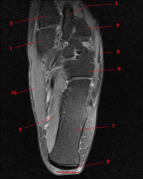
Near normal foot mri for reference. • muscle edema is seen secondary to multiple etiologies including trauma, infectious and inflammatory processes, autoimmune disorders, neoplasms, and denervation injuries • on mri muscle edema is characterized by increase in free water within the muscle • muscle edema is seen on mri as increased signal on fluid sensitive sequences t2 fs Magnetic resonance imaging (mri) is the modality of choice in diagnosing accessory muscles, delineating their relationship to adjacent structures, and differentiating them from soft tissue tumors. Those fibers of the most medial and largest belly are… The flexor digitorum brevis muscle lies immediately superior to the plantar aponeurosis and inferior to the tendons of the flexor digitorum longus in the sole of the foot.
Adductor hallucis is anatomically located in the central compartment of foot, but the muscle is functionally grouped with the medial plantar muscles of foot because it acts on the great toe (hallux).
This ensures anyone who will benefit from an mri to fully heal their pain can have one at an affordable cost. With a muscle injury, for example, mri images often show a bright signal indicating that there is more water in the muscle, which is a sign of injury. Mri is the choice of modality for further imaging the ankle and foot after obtaining initial radiographs. Mri is an ideal method for identifying areas of muscle atrophy and fatty infiltration. Routine ankle magnetic resonance imaging (mri) tests involve taking images of the foot and ankle in the axial, coronal, and sagittal planes parallel to the tabletop(2). The muscles lie within a flat fascia on the dorsum of the foot (fascia dorsalis pedis) and are innervated by the deep fibular or peroneal nerve. Denervation changes in muscles early. 23,25 mri at the level of the malleolus demonstrates the muscle as. In the foot and ankle many accessory ossicles can be seen. In addition, an image of all the muscles of the back and plantar part of the foot, all tendons and tendon ligaments, blood vessels and nerves are obtained. Muscle anatomy trivia 12 photos of the muscle anatomy trivia muscle anatomy trivia, human muscles, muscle anatomy trivia The adductor hallucis has two heads: Related posts of foot muscle anatomy mri muscle anatomy trivia.
Call or request an appointment online today and get your mri when it's convenient for you This imaging technique assesses the ligaments and tendons, neurovascular structures (tarsal tunnel and plantar fascia), and the osseous structures(19). The adductor hallucis has two heads: Anatomical structures of the ankle and foot and specific regions (major joints) are visible as dynamic labeled images. Magnetic resonance imaging (mri) is the modality of choice in diagnosing accessory muscles, delineating their relationship to adjacent structures, and differentiating them from soft tissue tumors.

The flexor digitorum brevis muscle lies immediately superior to the plantar aponeurosis and inferior to the tendons of the flexor digitorum longus in the sole of the foot.
Call or request an appointment online today and get your mri when it's convenient for you The deformity of the foot with abnormal pressure distribution on the plantar surface coupled with reduced or loss of sensation, makes the foot. Case contributed by dr andrew dixon. With a muscle injury, for example, mri images often show a bright signal indicating that there is more water in the muscle, which is a sign of injury. Magnetic resonance imaging (mri) is the modality of choice in diagnosing accessory muscles, delineating their relationship to adjacent structures, and differentiating them from soft tissue tumors. Routine ankle magnetic resonance imaging (mri) tests involve taking images of the foot and ankle in the axial, coronal, and sagittal planes parallel to the tabletop(2). Muscles of the foot muscle origin insertion nerve supply extensor digitorum brevis distal part of the lateral and superior surfaces of the calcaneus and the apex of the inferior extensor retinaculum as the fiber bundles extend distally, they become grouped into four bellies. The aim of this review is to provide the reader with a comprehensive overview of the magnetic resonance imaging (mri) characteristics of the most common benign and malignant soft tissue neoplasms which occur around the foot and ankle. Ultrasonography (us) affords high spatial resolution of muscle but is less sensitive than magnetic resonance (mr) imaging for mild edema and early myopathy. In addition, an image of all the muscles of the back and plantar part of the foot, all tendons and tendon ligaments, blood vessels and nerves are obtained. Accessory muscles are isointense to skeletal muscle on all pulse sequences, and can insert by fleshy muscular or tendinous insertions. The adductor hallucis has two heads: The peroneus quartus muscle is more common, presenting in 13% to 22% of the population.


Komentar
Posting Komentar